Introduction
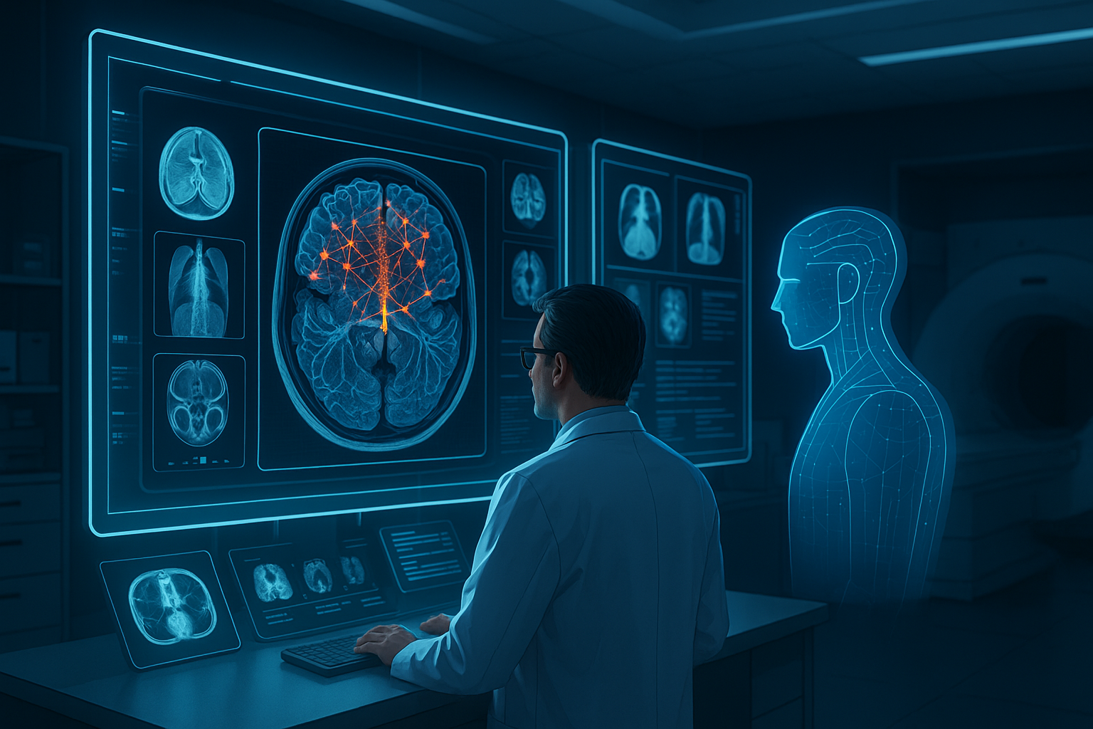
Radiology plays a crucial role in modern healthcare by using imaging techniques like X-rays, CT scans, and MRIs to detect and diagnose diseases. These tools allow doctors to see inside the human body without the need for surgery, making diagnosis safer and faster. However, reviewing thousands of images every day is time-consuming and can sometimes lead to mistakes due to human fatigue or oversight. That’s where Artificial Intelligence (AI) comes in.
AI is now making a big impact in radiology by helping doctors work more quickly and accurately. Two powerful types of AI—Deep Learning (DL) and Natural Language Processing (NLP)—are transforming the field. Deep learning focuses on understanding image data, while NLP helps make sense of written reports and doctors’ notes. Together, they allow computers to help label medical images, write reports, and even suggest possible diagnoses.
This article explores how deep learning and NLP are working together to make radiology smarter, faster, and more reliable.
The Importance of Medical Image Annotation
What is Medical Image Annotation?
Medical image annotation is the process of labeling specific parts of a medical image to show important information. For example, a radiologist might draw a circle around a tumor in an MRI scan or point out signs of pneumonia in a chest X-ray. These annotations help teach AI systems how to recognize diseases and other conditions in future images. Without labeled examples, AI wouldn’t know what to look for or how to interpret what it sees.
Annotations are not only useful for training AI but also for helping doctors during diagnosis. When an AI system marks a suspicious area, it acts as a second opinion, guiding doctors to double-check regions they might have overlooked. This leads to more accurate and faster decisions.
Challenges in Traditional Annotation
Despite its importance, annotating medical images by hand comes with many difficulties:
Takes a Lot of Time: Doctors often spend hours labeling images, especially when datasets contain thousands of files. This takes away time they could spend on patient care.
Different Opinions: Even expert radiologists may disagree on what an image shows, leading to inconsistencies in annotations.
Not Enough Experts: In many parts of the world, there are too few trained radiologists. This shortage slows down diagnosis and treatment.
Too Much Data: Hospitals and clinics generate massive amounts of imaging data every day—far more than humans can handle alone.
These issues show why automation is needed. AI offers a way to speed up the annotation process and make it more consistent.

The Emergence of Deep Learning in Radiology
What is Deep Learning?
Deep learning is a form of AI that uses computer models inspired by the human brain. These models are made of layers of “neurons” that process information step by step. The deeper the network (meaning the more layers it has), the better it can learn complex features.
One special type of deep learning called Convolutional Neural Networks (CNNs) is especially good at working with images. CNNs can learn to spot features like shapes, edges, and textures that are common in medical images. This makes them perfect for tasks like finding tumors or broken bones.

How Deep Learning is Used in Radiology
Deep learning models are already being used in hospitals and research labs for a wide variety of tasks:
-
Finding Problems: CNNs can detect abnormalities like cancerous tumors, fractures, or lung infections with high accuracy.
-
Drawing Boundaries: AI can outline organs, blood vessels, or disease regions to help doctors focus on important areas.
-
Sorting Images: AI can sort through huge collections of images and flag the ones that may show signs of disease.
-
Matching Images: Some models compare scans taken at different times to see how a disease is progressing or healing.
By automating these tasks, deep learning allows radiologists to focus on final decisions instead of time-consuming analysis.
Popular Deep Learning Models
Several deep learning models have become especially important in medical imaging:
-
U-Net: Designed for biomedical image segmentation, U-Net is great at outlining structures like organs or tumors.
-
ResNet (Residual Network): Enables the training of very deep models without losing earlier information.
-
DenseNet: Improves learning by connecting every layer to every other layer, leading to more accurate predictions.
-
YOLO (You Only Look Once) and Faster R-CNN: These models are fast and precise, making them useful for detecting diseases in real time.

The Role of Natural Language Processing in Radiology
What is NLP?
Natural Language Processing (NLP) is a type of AI that helps computers understand and generate human language. In radiology, NLP can read doctors’ notes, clinical summaries, and imaging reports. It turns this unstructured text into data that AI can understand and use for decision-making or training.
For example, NLP can read a report that says, “There is a small mass in the upper right lung,” and link it to the corresponding image, helping the system learn what that type of disease looks like.
How NLP Helps in Radiology
NLP makes radiology workflows more efficient in several ways:
Writing Reports: AI can generate first drafts of reports by summarizing what’s seen in the image.
Helping with Labels: NLP reads existing reports and extracts labels to use for AI training.
Finding Past Information: It enables quick searches through large archives of reports, helping doctors find similar past cases.
Supporting Decisions: NLP can suggest possible diagnoses or treatments based on prior reports and patient records.
Main NLP Techniques
Key NLP methods used in radiology include:
Named Entity Recognition (NER): Identifies important terms in a report, like diseases, organs, or medications.
Relation Extraction: Figures out relationships between entities—for instance, connecting a “tumor” with its location, such as “left lung.”
Transformer Models: Tools like BERT and GPT can understand complex language patterns and generate text that sounds natural and informative.

How Deep Learning and NLP Work Together
Learning from Both Images and Text
The real power of AI in radiology comes when deep learning and NLP are used together. Many medical images come with written reports, and combining these two data sources creates a more complete learning system.
For example, NLP can extract disease labels from reports, which are then used to train deep learning models on the matching images. Over time, the system gets better at spotting similar issues in new scans.
Better Ways to Add Labels
Here’s how the process usually works:
NLP reads past reports and finds useful labels.
Deep learning uses these labels to learn how diseases appear in images.
The AI model predicts labels for new images.
Doctors review these predictions and make corrections if needed.
This process is called “semi-automated labeling” and helps build large, high-quality datasets faster and with less effort.
Example: Reading Chest X-Rays
A good example is using AI to diagnose pneumonia from chest X-rays:
NLP scans past reports for mentions of pneumonia.
Deep learning models train on images labeled with pneumonia.
When a new X-ray comes in, the model predicts if pneumonia is present and even drafts a report for the doctor to edit.

Data Sources and Annotation Tools
Public Image Collections
To train AI models in radiology, researchers and developers need large amounts of labeled medical images. Fortunately, several public datasets are available that include images along with expert annotations or associated reports. These datasets help in training, testing, and benchmarking AI systems.
NIH ChestX-ray14: One of the most widely used datasets, it contains over 100,000 chest X-rays from more than 30,000 patients. It includes labels for 14 different diseases, such as pneumonia, cardiomegaly, and pneumothorax, extracted using NLP techniques.
MIMIC-CXR: Released by MIT, this dataset includes over 370,000 chest X-rays along with corresponding radiology reports. The reports are de-identified to protect patient privacy and are an excellent resource for combining NLP and imaging.
RSNA Pneumonia Detection Challenge Dataset: Created for a Kaggle competition, this set includes annotations from radiologists that highlight areas with pneumonia in pediatric chest X-rays.
VinDr-CXR: A high-quality dataset from Vietnam, annotated by experienced radiologists and designed to address the shortage of annotated medical data in Southeast Asia. It includes over 18,000 images.
These datasets allow AI systems to learn from real-world examples and improve performance across diverse populations and imaging conditions.

Tools for Labeling Images
Labeling tools are essential for creating training data. They allow radiologists and technicians to annotate images efficiently and consistently. Here are some commonly used tools in the field:
SO Development
Overview
SO Development is redefining the medical data collection space by leveraging AI-driven platforms tailored for large-scale, high-quality datasets.

Labelbox:
A user-friendly platform that allows annotation of medical images using bounding boxes, polygons, and segmentation tools. It also supports team collaboration and quality checks.
MD.ai
Specifically designed for the medical field, MD.ai offers tools for labeling DICOM images and linking them with NLP-processed reports.
V7 Darwin
Offers advanced automation features, like auto-labeling and AI-assisted segmentation. It integrates easily with deep learning frameworks.
MONAI Label
An open-source tool from Project MONAI designed for medical imaging. It integrates with medical viewers and allows AI-assisted interactive labeling.
These platforms not only speed up the annotation process but also help manage complex workflows, ensuring data quality and security.
How AI Models Are Trained
Training an AI model for radiology involves several key steps, each of which must be handled with care to ensure accuracy and safety.
Preparing the Data
Before training can begin, the image and text data must be cleaned and standardized:
Image Preprocessing: This may include adjusting brightness and contrast, normalizing resolution, and converting images to a consistent format. Often, DICOM files are converted to PNG or JPEG for easier processing.
Text Cleaning: Radiology reports may contain typos, abbreviations, or inconsistent formatting. NLP preprocessing involves tokenizing text, correcting misspellings, and standardizing terminology.
Data Matching: Images are linked to their corresponding reports using patient IDs or study IDs, ensuring that each image has the correct labels.
Adding Annotations
There are three common methods for obtaining annotations:
Manual Labeling: Radiologists manually mark regions of interest on the images, which is time-consuming but highly accurate.
NLP-Based Label Extraction: Algorithms extract labels from reports using NLP techniques. This method is faster but can introduce some errors.
Consensus Annotation: A combination of multiple human annotations is used to create a “gold standard” label, often achieved through majority voting or expert review.
The quality of these labels directly affects how well the AI model will perform.
Training the AI
Once the data is ready, training begins:
Model Selection: Depending on the task (classification, segmentation, or detection), different models are chosen.
Loss Functions: Special functions are used to measure how well the model is doing. Common loss functions include cross-entropy for classification and Dice loss for segmentation.
Data Augmentation: Techniques like rotating, flipping, and zooming images are used to create more training examples and prevent overfitting.
Hyperparameter Tuning: Settings like learning rate, batch size, and number of epochs are adjusted to optimize performance.
This process may take hours or even days, depending on the size of the dataset and the complexity of the model.
Testing the Model
Once trained, the model must be tested thoroughly:
Holdout Testing: A portion of the dataset is set aside for testing and not used during training.
Cross-Validation: Data is divided into folds to ensure the model performs well across different samples.
Metrics: Accuracy, precision, recall, F1 score, and AUC-ROC are commonly used to evaluate performance.
Radiologist Agreement: The AI’s predictions are compared with those of expert radiologists to ensure clinical reliability.
Only after passing these tests can a model be considered ready for deployment in clinical settings.
How AI Helps in Hospitals
AI tools are being integrated into hospital systems to assist radiologists and improve workflows. These tools are designed not to replace doctors, but to support them by taking over repetitive tasks and highlighting important findings.
Sorting Urgent Cases
AI systems can scan incoming images and flag those that show signs of serious conditions—such as internal bleeding or collapsed lungs. This helps doctors prioritize cases and respond more quickly in emergencies.
For example, a CT scan showing a potential stroke could be automatically placed at the top of the review queue. In life-threatening situations, even a few minutes can make a big difference.
Writing Reports
AI can draft radiology reports by analyzing images and generating sentences that describe the findings. These drafts are reviewed and edited by doctors, saving them time while maintaining quality.
This feature is especially helpful in busy hospitals, where radiologists may need to write dozens of reports a day. It reduces burnout and increases consistency across reports.
Checking Quality
AI can act as a second reader, reviewing images after a human radiologist has completed their analysis. This “double-check” helps catch missed abnormalities and improves patient safety.
In some hospitals, AI systems have helped reduce error rates by identifying missed fractures, lung nodules, or subtle changes that were initially overlooked.
Working Together
Studies have shown that radiologists who use AI tools perform better than either humans or AI alone. AI can point out patterns or highlight areas of concern, but the final judgment remains with the doctor.
This collaborative approach is often referred to as “human-in-the-loop” AI and represents the future of clinical decision-making.

Ethics and Safety Rules
As AI becomes more common in healthcare, it’s critical to ensure that its use is ethical, fair, and safe for all patients.
Keeping Data Private
Medical data is highly sensitive. Regulations such as:
HIPAA (U.S.)
GDPR (Europe)
require that patient information be protected. This includes removing names, addresses, and other personal identifiers before using data for AI training.
Hospitals must also ensure that data is stored securely and only accessible to authorized personnel.
Fairness and Bias
AI models can become biased if they are trained on data from only one type of patient population. For example, a model trained mostly on scans from adult patients might perform poorly on children or people from different ethnic backgrounds.
To avoid this, developers should:
Use diverse datasets
Monitor model performance across demographics
Regularly audit outcomes for fairness
Making AI Understandable
Doctors need to trust AI tools, which means the tools must be explainable. Techniques such as heatmaps or saliency maps can show which parts of an image influenced the AI’s decision.
Explainable AI helps radiologists understand how the model reached a conclusion, allowing them to verify or challenge its results.
Legal Approvals
Before AI tools can be used in real clinics, they must receive approval from regulatory bodies such as:
U.S. Food and Drug Administration (FDA)
European Medicines Agency (EMA)
China’s NMPA
Approval processes involve reviewing the model’s accuracy, safety, and usability in a clinical environment.

Challenges to Overcome
Despite its potential, AI in radiology still faces many challenges:
Bad Quality Images: Poor resolution or improperly scanned images can confuse AI models.
Limited Use Cases: Some AI tools only work well in the specific hospital or environment they were trained in.
Wrong Labels: NLP-based labels from reports can contain errors, especially if the original report was vague or inconsistent.
High Costs: Training large models requires powerful hardware, specialized staff, and long development times.
Researchers are actively working on solutions, such as transfer learning and lightweight models, to make AI more accessible and robust.

What’s Next for AI in Radiology?
The future of AI in radiology is full of possibilities. As the technology improves, we can expect several exciting developments:
Teamwork Across Hospitals
Federated learning allows hospitals to train shared AI models without sharing raw data. Instead, the model learns locally and only shares updates, protecting patient privacy while improving performance.
This approach helps build powerful models from global datasets without violating data protection laws.
Learning Without Labels
Self-supervised and unsupervised learning techniques are being developed to allow AI models to learn from unannotated data. This greatly reduces the need for manual labeling and speeds up development.
For instance, a model could learn to group similar images together even if it doesn’t know what disease is present.
New Tests and Standards
To ensure AI systems are safe and effective, new international standards and benchmarking tests are being created. These include challenges, public competitions, and shared testing platforms.
Common testing frameworks will allow fair comparison of models and promote transparency in performance claims.
Personalized Medicine
In the future, AI could combine imaging data with other health information—like genetic tests, lifestyle factors, and medical history—to provide personalized recommendations.
This could lead to earlier detection, more accurate diagnoses, and treatments tailored to each patient.
Better Integration
As AI tools mature, they will be better integrated into existing hospital systems. This means faster deployment, real-time results, and seamless communication with electronic health records (EHRs).
Doctors will be able to access AI insights within the same software they already use, making the technology more practical and widely adopted.

Conclusion
Artificial intelligence is revolutionizing the field of radiology. With the help of deep learning and natural language processing, machines can now analyze medical images, extract meaningful patterns, and assist in report writing. These tools are not replacing radiologists, but empowering them to work faster and with greater accuracy.
From detecting diseases to prioritizing urgent cases, AI offers solutions that improve patient care and hospital efficiency. As the technology evolves, it will play an even bigger role—enabling personalized medicine, enhancing collaboration across institutions, and setting new standards in healthcare innovation.
The future of radiology is not only digital but intelligent. And with AI as a trusted partner, radiologists are better equipped than ever to see what matters most.